- Visibility 14 Views
- Downloads 2 Downloads
- DOI 10.18231/j.jdp.2022.036
-
CrossMark

Effectiveness of resin infiltration method on white spot lesions post orthodontic treatment - A systematic review
- Author Details:
-
 Sushmita Batni Rao *
-
Taher Manasawala
-
Devashree Mujumdar
Introduction
Orthodontic treatment aims to improve both facial and functional aesthetics that benefits oral and overall health of a person. However, any treatment could be related to few complications but can be avoided by altering the usual lifestyle. The most known risk that can be developed during or after orthodontic treatment is the demineralization of enamel surface also known as White Spot Lesions which are known to occur despite of having many treatment alternatives.[1]
The term "white spot lesion" (WSL) as defined by Fejerskov et al. is "the first sign of caries lesion on enamel that can be detected with the naked eye" and is used along with the terms "initial" or "incipient" lesions. [2] They are usually seen around the periphery of the brackets as small lines but may also be seen as large areas of decalcifications is few cases. [3]
White spot lesions usually occur as a sign of initial decay showing a disproportion between demineralization and remineralization of the enamel surface. WSLs can be caused due to bacterial invasion due to their acid production since it is said to contain sugars which leads to destruction of the surface.[4]
Usually WSLs have an opaque, white and a chalky appearance which is due to an optical phenomenon which is caused due to loss of minerals in the surface and subsurface areas of the enamel.[5]
Many studies have stated that the overall prevalence of the damage to the enamel surface after orthodontic treatment ranges between 2-96% as compared to controls and the most affected teeth are said to be molars, mandibular canines, premolars, and maxillary lateral incisors.[6]
Considering the development of these lesions, orthodontists face many challenges owing to the early diagnosis and treatment of WSLs, and this requires complete knowledge about the disease and factors causing it which are unique with every patient. These problems should be brought into evaluation at a very early stage to minimize the risk of persistent tooth decay and its effects. [1]
Therefore, it would be very useful for the orthodontist to take up methods for prevention and control the formation of WSLs if they are detected in their early stages during orthodontic treatment. [7]
Many studies have been carried out in relation to the effectiveness of resin infiltration for cavitated enamel lesions but not many studies signify its effect after orthodontic debonding. Therefore, the aim of this systematic review was to evaluate the effectiveness of resin infiltration method on white spot lesions post orthodontic treatment.
Materials and Methods
Protocol
This systematic review was performed based on the given PRISMA statement guidelines. [8]
Focus question
Effectiveness of resin infiltration method on white spot lesions post orthodontic treatment.
Study design
Randomized Clinical Trials (RCTs) or Controlled Clinical Trials (CCTs) and In-Vivo studies.
Search strategy
Four electronic databases (PubMed, Google Scholar, Embase and Cochrane Library Database) were searched from 2009 to 31st October 2019 for original studies of the required criteria. Only articles in English were reviewed.
The search terms under MeSH heading were “White Spot Lesions” AND “Resin Infiltration” AND “Orthodontic Debonding”. Papers with ‘in-vitro’ and ‘in- situ’ studies with artificial White Spot Lesions were not taken into consideration. The reference lists of the accepted papers were hand-searched for additional literature. Key data of the accepted studies were independently reviewed by two reviewers.
Inclusion criteria
The PICO was constructed as follows:
P- Young adults and adolescents aged less than 30 years with white spot lesions seen within 3-4 months post orthodontic treatment.
I- Any intervention except for laminates or veneers to converse post-orthodontic lesions or improving esthetics.
C- No treatment or Placebo.
O- Effectiveness of the resin used and esthetics in terms of color masking.
Exclusion criteria
Studies comparing the effectiveness of resin infiltration with other treatment modalities.
Studies performed without using resin infiltration technique.
Studies other than RCT’s or In- Vivo.
Risk assessment
Two review authors (VS and SB) independently assessed the risk of bias for the selected trials using Cochrane’s tool for assessing risk of bias, as described in section 8.5 of the Cochrane Handbook for Systematic Reviews of Interventions. [9]
We assessed the following domains as being at low, high or unclear risk of bias:
Random sequence generation (selection bias)
Allocation concealment (selection bias)
Blinding of participants and personnel (performance bias)
Blinding of outcome assessors (detection bias)
Incomplete outcome data addressed (attrition bias)
Selective outcome reporting (reporting bias)
Other bias.
Overall risk of bias
The overall risk of bias for all the studies in the systematic review is unclear.
Results
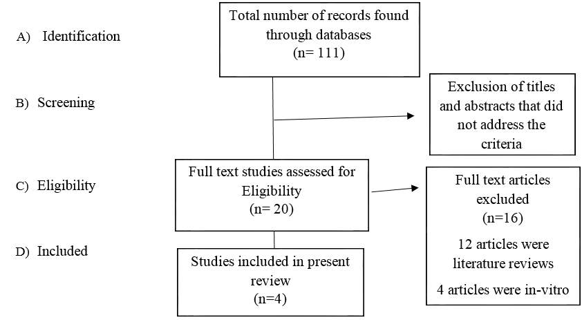
A total of 111 records were found through all the databases out of which, a total of 12 articles were literature reviews relevant to the study and only 4 articles[10], [11], [12], [13] were RCTs which met the inclusion criteria ([Figure 1]).
Two authors independently extracted information regarding methods, participants, interventions, outcome measures and results and the risk of bias for each study was evaluated ([Table 1]).
|
Study |
Random Sequence Generation |
Allocation Concealment |
Blinding of participants and Personnel |
Blinding of outcome assessment |
Incomplete Outcome Data |
Selective Reporting |
Other Bias |
|
Michael Knosel et al. [10] |
? |
+ |
? |
? |
? |
? |
? |
|
Eckstein et al. [11] |
? |
+ |
? |
? |
? |
? |
? |
|
Senestraro et al. [12] |
- |
- |
? |
? |
? |
? |
? |
|
Knosel et al. [13] |
? |
- |
|
? |
? |
? |
? |
Discussion
The development of WSLs is seen to be a most common secondary response to orthodontic treatment and many treatment options are available in the market today. [14]
The treatment of WSLs using resin infiltration technique has been shown to have positive results in its effectiveness to be able to camouflage with the adjacent enamel based on the researched studies. But the long-term persistence of the technique remains questionable to both patients as well as clinicians. [3], [15]
This systematic review is unable to provide adequate scientific support because it includes only in-vivo and Randomized Clinical Trials (RCTs) of resin infiltration on white spot lesions (WSL) developed post-debonding compared to sound enamel from the available literature.
The first study included the re-assessment of long-term camouflage effects of resin infiltration (ICON) on white spot lesions (WSL) compared to sound enamel which was achieved in a previous trial. The subjects who had received resin infiltration in the previous trial after fixed orthodontic treatment were reassessed for the effectiveness of the resin. The results achieved by this technique were shown to be effective in the hue and croma, and no changes were observed over the last 24 months.
The second study was an in-vivo study assessing the camouflage effects by concealment of post-orthodontic white-spot lesions to sound adjacent enamel achieved over 12 months with resin infiltration (ICON). Out of 20 subjects selected, only 9 subjects were available at the end of 12 months for assessment. The results after a 12-month analysis stated that there was a decrease in the color and lightness of WSLs when compared to adjacent sound enamel which indicated its assimilation.
The 3rd and 4th studies were Randomized Clinical Trials (RCTs) where the subjects selected were participants who had completed orthodontic treatment to assess changes in the appearance of white-spot lesions (WSLs) that were treated with resin infiltration. The teeth under the treatment group were evaluated for changes using photographs and a visual analog scale.
There was a significant improvement in the clinical appearance of white spot lesions which had developed during orthodontic treatment seen in 8 months and 12 months respectively.
Conclusion
Based on currently available literature and after viewing them systematically, the following can be concluded:
Long term improvement in the aesthetic appearance of WSLs after orthodontic treatment can be considered suitable when compared to the color of adjacent enamel surface.
ICON technique is said to have an improvement on the optical appearance of WSLs.
There are no alterations observed with respect to the colour and lightness of the adjacent sound enamel.
Consistent stability over a period of 12 months as seen in all 4 studies can be achieved using the ICON for masking WSLs with adjacent sound enamel.
Finally, the recent advancement of resin infiltration method for masking White Spot Lesions has been proved successful and better as compared to other traditional techniques.
However, due to a limited number of studied performed and risk of bias, more studies should be undertaken to assess the effectiveness of resin infiltration method on white spot lesions post orthodontic treatment.
Source of Funding
No financial support was received for the work within this manuscript.
Conflict of Interest
None declared.
References
- H Aghoutan, S Alami, F El Quars, S Diouny, F Bourzgui. White Spots Lesions in Orthodontic Treatment and Fluoride-Clinical Evidence. Emerg Trends Oral Health Sci Dent 2015. [Google Scholar]
- E Zabokova-Bilbilova, L Popovska, B Kapusevska, E Stefanovska. White spot lesions: prevention and management during the orthodontic treatment. Pril (Makedon Akad Nauk Umet Odd Med Nauki) 2014. [Google Scholar] [Crossref]
- B Ogaard, G Rølla, J Arends. Orthodontic appliances and enamel demineralization. Part 1. Lesion development. Am J Orthod Dentofacial Orthop 1988. [Google Scholar]
- E Tufekci, JS Dixon, JC Gunsolley, SJ Lindauer. Prevalence of white spot lesions during orthodontic treatment with fixed appliances. Angle Orthod 2011. [Google Scholar]
- GC Heymann, D Grauer. A contemporary review of white spot lesions in orthodontics. J Esthet Restor Dent 2013. [Google Scholar]
- K Lopatiene, M Borisovaite, E Lapenaite. Prevention and Treatment of White Spot Lesions During and After Treatment with Fixed Orthodontic Appliances: a Systematic Literature Review. J Oral Maxillofac Res 2016. [Google Scholar]
- KC Julien, PH Buschang, PM Campbell. Prevalence of white spot lesion formation during orthodontic treatment. Angle Orthod 2013. [Google Scholar]
- A Liberati, DG Altman, J Tetzlaff, C Mulrow, PC Gøtzsche, JP Ioannidis. The PRISMA statement for reporting systematic reviews and meta-analyses of studies that evaluate health care interventions: explanation and elaboration. Ann Intern Med 2009. [Google Scholar]
- J P Higgins, D G Altman, P C Gøtzsche, P Jüni, D Moher, A D Oxman. Cochrane Bias Methods Group; Cochrane Statistical Methods Group. The Cochrane Collaboration's tool for assessing risk of bias in randomised trials. BMJ 2011. [Google Scholar] [Crossref]
- M Knösel, A Eckstein, H J Helms. Long-term follow-up of camouflage effects following resin infiltration of post orthodontic white-spot lesions in vivo. Angle Orthod 2019. [Google Scholar]
- A Eckstein, HJ Helms, M Knösel. Camouflage effects following resin infiltration of postorthodontic white-spot lesions in vivo: One-year follow-up. Angle Orthod 2015. [Google Scholar]
- SV Senestraro, JJ Crowe, M Wang, A Vo, G Huang, J Ferracane. Minimally invasive resin infiltration of arrested white-spot lesions: a randomized clinical trial. J Am Dent Assoc 2013. [Google Scholar] [Crossref]
- M Knösel, A Eckstein, H J Helms. Durability of esthetic improvement following Icon resin infiltration of multibracket-induced white spot lesions compared with no therapy over 6 months: a single-center, split-mouth, randomized clinical trial. Am J Orthod Dentofacial Orthop 2013. [Google Scholar]
- M Sonesson, F Bergstrand, S Gizani, S Twetman. Management of post-orthodontic white spot lesions: an updated systematic review. Eur J Orthod 2017. [Google Scholar]
- C Rocha Gomes Torres, AB Borges, LM Torres, IS Gomes, RS De Oliveira. Effect of caries infiltration technique and fluoride therapy on the colour masking of white spot lesions. J Dent 2011. [Google Scholar]